. 2020 Sep 24;11(41):11322–11330. doi: 10.1039/d0sc04520a

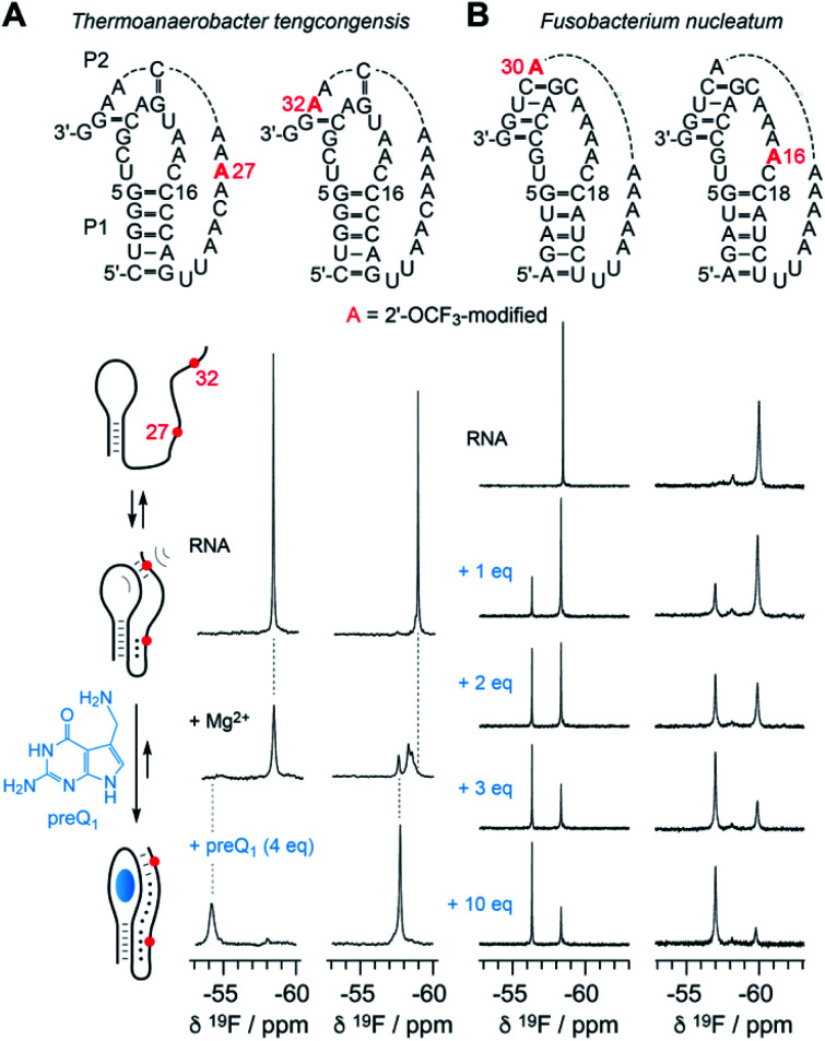
Fig. 6. NMR spectroscopic analysis of RNA-ligand binding. (A) Secondary structure of Thermoanaerobacter tengcongensis preQ1-I riboswitch with 2′-OCF3 position indicated in red (top); schematic folding model (left) of aptamer-assisted RNA pseudoknot formation, and stabilization through binding of the small molecule 7-aminomethyl-7-deazaguanine (preQ1) indicated in blue; 19F NMR (564 MHz) spectra of RNA in response to Mg2+ and preQ1 addition (right). Conditions: c(RNA) = 0.14 mM, 15 mM sodium cacodylate, 25 mM NaCl, pH 6.5, 297 K; additions: c(Mg2+) = 2.0 mM; followed by c(preQ1) equivalents as indicated. (B) Secondary structure of Fusobacterium nucleatum preQ1-I riboswitch with two distinct 2′-OCF3 positions in red (top). 19F NMR (564 MHz) spectra of RNA in response to preQ1 titrations (right). Conditions: c(RNA) = 0.3 mM, 25 mM sodium cacodylate, pH 6.5, 298 K; additions: c(Mg2+) = 2.0 mM; followed by c(preQ1) equivalents as indicated.

Fig. 6