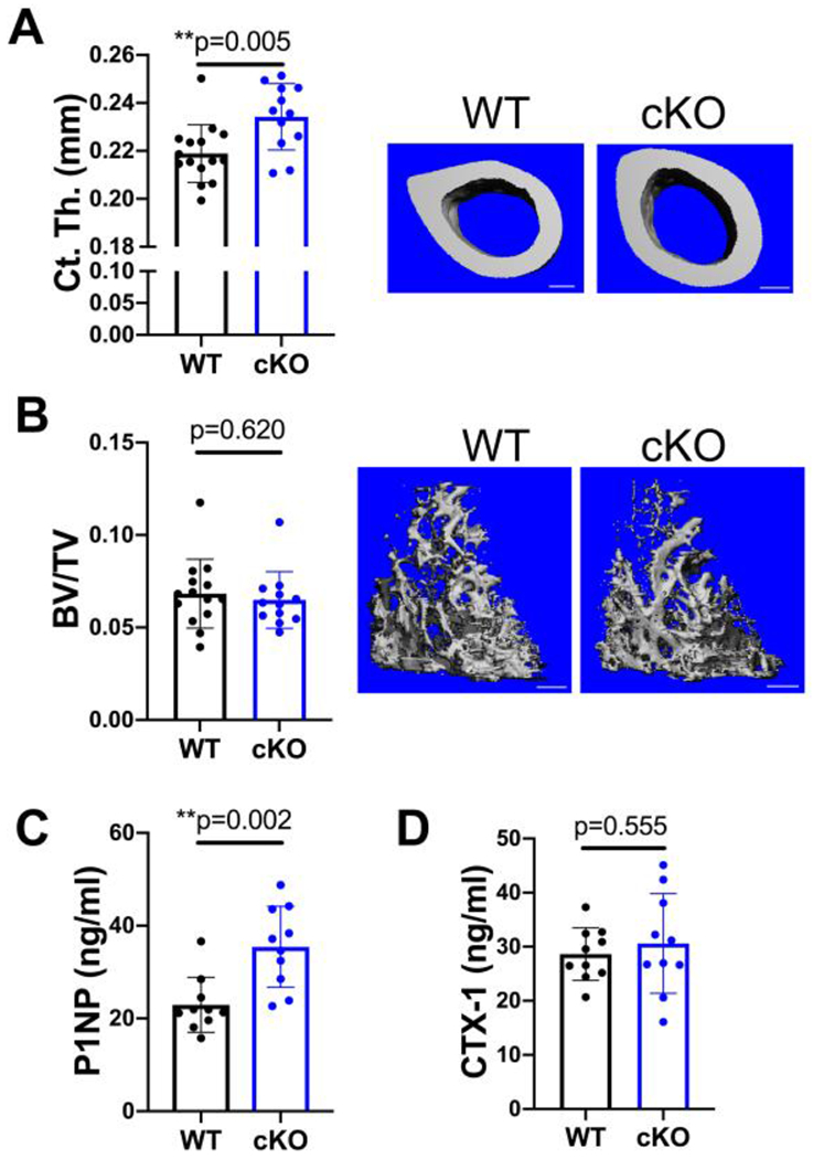
Figure 2.

Only cortical bone is increased by MFN2 deficiency at peak bone mass in females. A,B) MicroCT analysis of the distal femur shows that cortical thickness (Ct.Th A) is increased significantly while trabecular bone volume fraction is similar (BV/TV, B) at 18 weeks of age. Representative images with scale bars = 400 μm. C) The serum marker of bone formation, P1NP, is increased in MFN2 cKO 18 week females. D) CTX-1 remains unchanged at this age. Unpaired t-tests, n=10.
