. 2020 Sep 24;11(41):11266–11273. doi: 10.1039/d0sc04849f

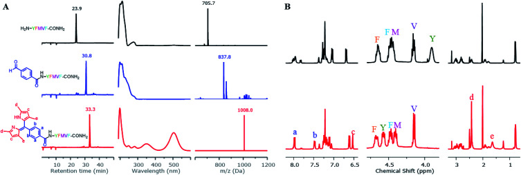
Fig. 2. Direct construction of dipyrrin on resin–bound Pep1. (A) The crude samples (resin) were taken and cleaved, with the resulting solutions being monitored by HPLC and ESI-MS. From top to bottom: unconjugated peptide (black), aldehyde–peptide conjugate (blue), dipyrrin–peptide conjugate DP1–Pep1 (red); from left to right: their HPLC chromatographs, UV-Vis spectra corresponding to the peak on HPLC, and ESI-MS spectra. (B) Comparisons of 1H-NMR spectra of Pep1 and DP1–Pep1: four protons from the aldehyde building block (a and b) and two protons from the pyrrole (c) were found in the aromatic region; the α-H of tyrosine (Y, near the N-terminal) shifts largely after conjugation; 12 protons from the four methyl groups of dipyrrin (d and e) were found at 2.4 and 1.7 ppm.

Fig. 2