. 2020 Sep 24;11(41):11266–11273. doi: 10.1039/d0sc04849f

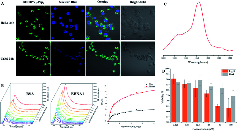
Fig. 6. Imaging of fluorescent peptides. (A) Confocal imaging of BODIPY1–Pep4 in HeLa and C666 cell lines. The green fluorescence signal was detected in the nucleus of C666 (EBNA+) but not in the nucleus of HeLa (EBNA−) cells. (B) Fluorescence titration of BSA (left) and EBNA1 (middle) proteins, both at 1.2 μM, with BODIPY1–Pep4. A nearly 5-times enhancement over the course of the titration with EBNA1 was observed (right). (C) Singlet oxygen signal illustrating the potential of BODIPY1–Pep4 as a PDT agent. (D). Comparison of dark/light cytotoxicity of BODIPY1–Pep4 towards C666 cell line. Light toxicity was significantly higher than the dark toxicity. Error bars represent the standard derivation of the mean.

Fig. 6