. 2020 Oct 12;11(44):12212–12220. doi: 10.1039/d0sc04751a

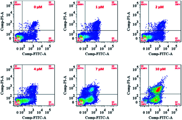
Fig. 5. Flow cytometry analysis of MCF-7 cell apoptosis after treatment with Ir-Rho-G2 at different concentrations. MCF-7 cells were seeded at 8 × 10³ to 1 ×10⁴ cells per well in 96 well plates then incubated for 24 h in 5% CO2 at 37 °C. The solutions of the complexes (10 mM in DMSO) were diluted to appropriate concentrations with the complete medium and added to the cells to give final complexes concentrations (1, 2, 5, 10 μM). After incubation for 2 h, the well plate was irradiated with 11 W lamp for 30 min. After the irradiation, the cells were again incubated for 4 h, after which the cell apoptosis was determined by standard flow cytometry analysis approach.

Fig. 5