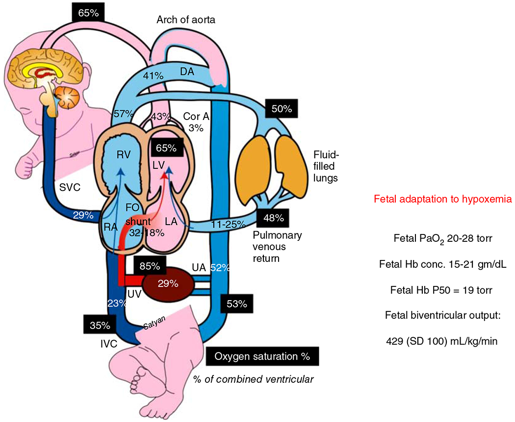
Figure 1.

Adaptation of the fetus to low oxygen environment present in utero. Fetal oxygen supply is provided by maternal blood bathing the chorionic villi in the placenta. The umbilical vein carries the oxygenated blood to inferior vena cava (IVC) and eventually across the foramen ovale to left atrium and left ventricle to be pumped into coronary and cerebral circulations. The oxygen saturation of fetal blood in different sites are indicated by dark shaded boxes. The percent of cardiac output distributed to each organ is indicated in plain text. The mean values for fetal biventricular output, range of normal Hb concentrations and fetal PaO2 are indicated in the text box to the right, along with the HbP50 for fetal Hb (197, 257, 268). Reused, with permission, from Richard A. Polin and William W Fox, 2016, Fetal and Neonatal Physiology, Ed: Polin, Abman, Rowitch, Benitz and Fox, 5th edition, Lakshminrusimha and Steinhorn “Pathophysiology of PPHN,” pp 1576-1587. Copyright Satyan Lakshminrusimha.
