
Fig. 3. Gestational BPA exposure accelerates hypothalamic neurogenesis.
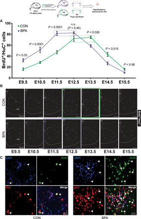
Sections of the tuberal hypothalamus were stained for BrdU+/HuC+ neurons born on each day during the window of hypothalamic neurogenesis (E9 to E15). BPA exposure resulted in more early-born neurons in this window and less late-born neurons. Quantification (A) (two-way ANOVA, P values compare treatments at each time point) and representative images (B) are shown, with peak neurogenesis indicated by green (CON) or purple (BPA) boxes. The third ventricle edge (3V), presumptive ventromedial hypothalamus (VMH), and the pial edge is shown in white dashed outline. Representative high-magnification images of dual-positive cells are shown within the VMH of CON and BPA mice (C). IHC, immunohistochemistry.