Fig. 5. Ex vivo hypothalamic NSPC culture elucidates mechanisms of BPA action.

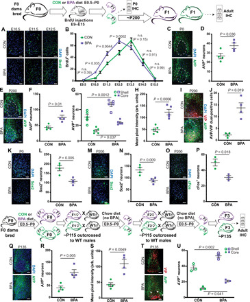
BPA treatment (10 nM) increased the number [representative images in (A) and quantified in (B)] and size [representative images in (A) and quantified in (C)] of primary neurospheres. BPA (10 nM) decreased the number [representative images in (D) and quantified in (E)] but increased the size [representative images in (D) and quantified in (F)] of secondary neurospheres. We observed significant reduction in sphere-forming cells in tertiary (G) and quaternary (H) cells but not in the fifth culturing (I). Differentiated 10 days in vitro (DIV) BPA-treated cells expressed higher NeuN [representative images in (J) and quantified in (K)] and lower Sox2 [representative images in (L) and quantified in (M)] than controls. Gestational BPA exposure increased primary neurosphere number without direct BPA treatment [graphical experimental explanation in (N) and quantification in (O)] even when BPA diet was only given in the past [quantification in (P)]. Use of fulvestrant (Fulv) and flutamide (Flut) indicates that dual ER/AR antagonism is required for phenotypic rescue of primary sphere number [representative images in (Q) and quantified in (R), ANOVA] and size [representative images in (Q) and quantified in (S), ANOVA]. Combined antagonism also reversed BPA effects on secondary sphere number [representative images in (T) and quantified in (U), ANOVA] and size [representative images in (T) and quantified in (V), ANOVA]. NS, neurospheres; n.s., no significance.
