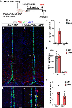
Fig. 2. Loss of Nfia/b/x selectively induces tanycyte proliferation in adult mice.

(A) Schematic for intracerebroventricular (i.c.v.) delivery of AAV-Cre and analysis of Nfia/b/x loss of function in P78 mice. (B) AAV-Cre induces Sun1-GFP expression in tanycytes in CAG-lsl-Sun1-GFP control mice at P78 [(b) inset shows alpha tanycytes]. (C) AAV-Cre induces proliferation in alpha tanycytes [shown in inset (c)] of Nfialox/lox;Nfiblox/lox;Nfixlox/lox;CAG-lsl-Sun1-GFP mice. (D) Quantification of GFP+/EdU+ cells in VZ and HP in NFI-deficient mice (n = 4 to 5 mice). (E) Number of GFP+ cells in VZ and HP in control and NFI-deficient mice. (F) Percentage of GFP+ VZ cells in the alpha tanycyte region labeled by Ki67 and EdU in NFI-deficient mice (n = 4 to 5 mice). Scale bars, 100 μm (B and C) and 25 μm (b and c).
