
Fig. 7. Nfia/b/x-deficient tanycytes mostly differentiate into neurons.
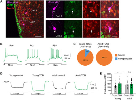
(A) Low- and high-magnification confocal images showing two biocytin-filled GFP+ recorded cells (white arrows) in an NFI TKO brain slice stained with NeuN. (B) Example responses of tanycyte-derived cells to depolarizing current steps. (C) The proportion of TDNs among tested tanycyte-derived cells in young (left) and adult (right) mice. (D) Representative average responses to hyperpolarizing current steps. (E) Summary graphs of input resistance (young control neurons, 18 cells from five mice, 1101 ± 89 MΩ; young TDNs, 17 cells from five mice, 1546 ± 176 MΩ; P = 0.0238, Mann-Whitney U test; adult control neurons, 16 cells from six mice, 1369 ± 132 MΩ; adult TDNs, 18 cells from six mice, 1477 ± 119 MΩ; P = 0.4777, Mann-Whitney U test). Scale bars, 50 μm (A, left) and 20 μm (A, right). *P < 0.05.