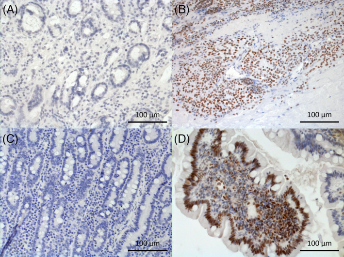
FIGURE 2.

Immunohistochemical staining of phosphorylated signal transducer and activator of pSTAT3 in duodenal mucosa of, A, negative control; B, positive control (mammary carcinoma); C, control (beagle) dog crypt region; D, SRE dog villus region; cell nuclei positive for pSTAT3 are stained in brown, negative ones are stained in blue with hematoxylin
