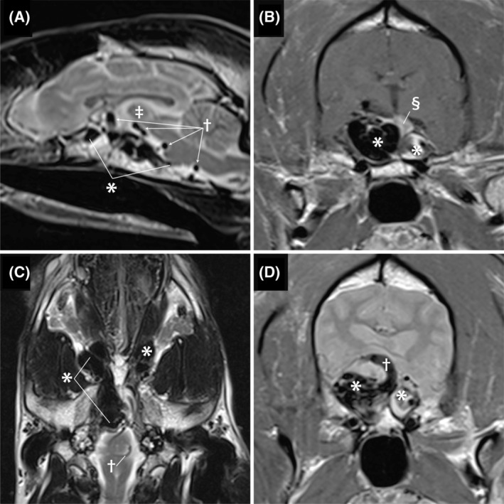
FIGURE 1.

Pretreatment of magnetic resonance images. Image A is a sagittal T2‐weighted image. The complex aggregation of hypoechoic structures along the ventral calvaria (*) represents the arteriovenous malformation (AVM). The AVM is causing a mass effect that is dorsally deviating the intrathalmic adhesion (double dagger). The round structures with a signal void indicated by the cross are the engorged serpentine basilar artery. Image B is a transverse post‐contrast T1‐weighted image. The large bilobed shape of the AVM is indicated by the *. The right side is larger and represents the confluence of multiple abnormal vessels including the cavernous sinus and the left side represents distention of the contralateral cavernous sinus. The contrast enhancing pituitary indicated by the double S symbol is dorsally displaced. Image C is a dorsal plane T2‐weighted image. The hypointense structures indicated by the * represent severe dilatation of the ophthalmic plexus that is worse on the right and the cause of exophthalmos. Image D Is a transverse plane image. The bilobed mass (*) causes dorsal elevation and compression of the thalmus. The dorsally curving vessel (cross) is the basilar artery connecting with the AVM
