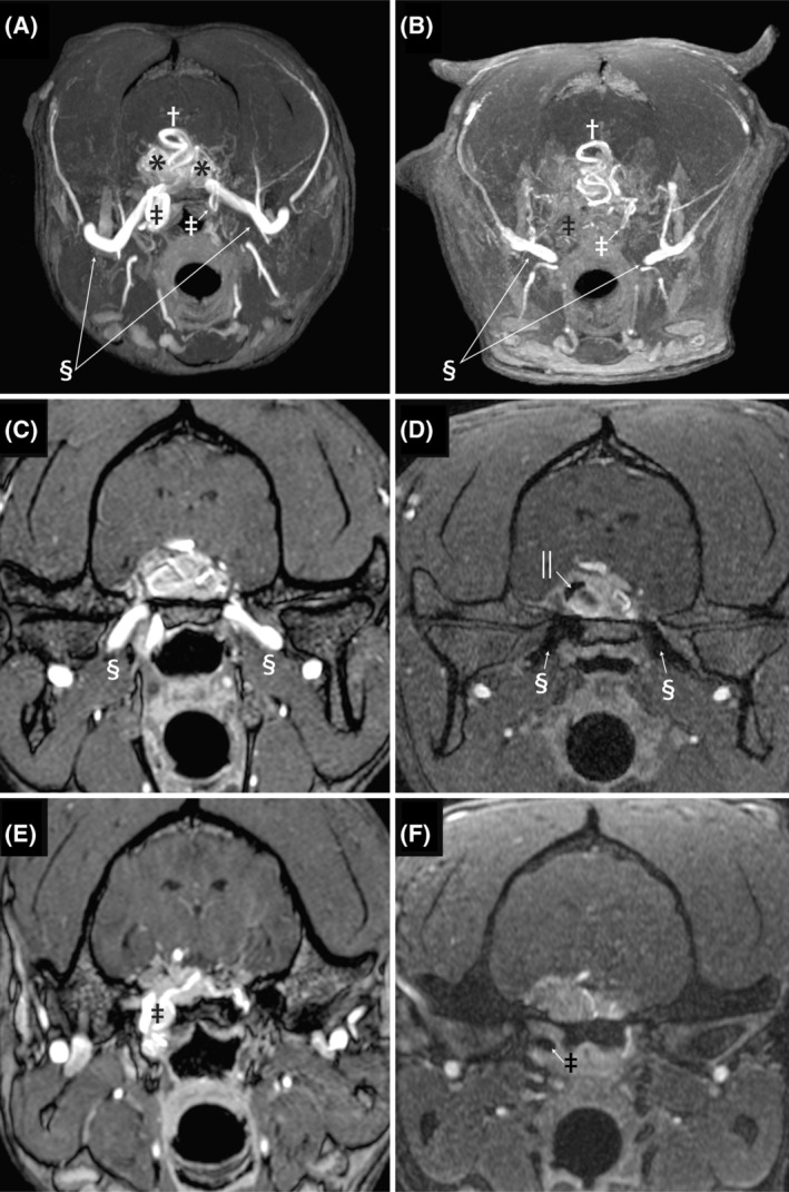
FIGURE 3.

Images A and B are pre‐ and post‐embolization transverse maximum intensity time‐of‐flight images. On image A, the maxillary arteries are clearly visible indicated by the double S symbol, and after embolization (B) both the left and right maxillary arteries abruptly truncate before entering the calvaria. The previously dilated and tortuous right‐sided internal carotid artery (black double dagger) at the level of the foramen lacerum is absent in the post‐embolization image, while the small normally sized left internal carotid artery (white double dagger) can be seen to take a hairpin loop at this level on both the pre‐ and post‐embolization images. Images C and D are time‐of‐flight images at the level of the oval foramen. The left and right maxillary arteries have flow present on the pre‐embolization images as indicated by the double S symbol. After embolization, both the maxillary arteries are occluded with hypointense material. Additional hypointense embolization material is evident within the AVM (||). Images E and F are transverse time‐of‐flight images at the level of the foramen lacerum. Image E is pre‐embolization and the dilated, tortuous internal carotid artery is evident (double dagger). Image F is post‐embolization and there is hypointense embolization material and no flow is identified in the internal carotid artery (double dagger) at that level
