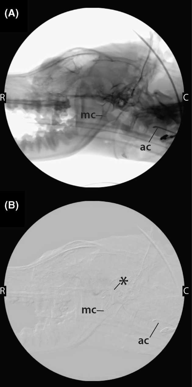
FIGURE 5.

Nonsubtracted (A) and subtracted (B) fluoroscopic images obtained with the dog in lateral recumbency during placement of instrumentation for the performance of liquid embolization. The 4Fr.‐angled catheter (ac) can be visualized in the maxillary artery, and a microcatheter (mc) has been coaxially introduced into the angled catheter and passed into the AVM nidus (*). C caudal; R, rostral
