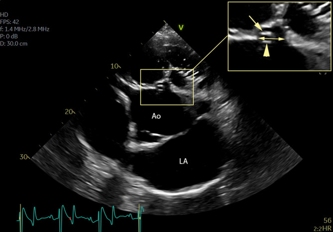
FIGURE 2.

Short‐axis view of the perimembranous ventricular septal defect, adjacent to the commissure of the right and noncoronary cusp, of the same horse as in Figure 1. The sinus of Valsalva (arrow) and opened aortic valve cusp (arrowhead) are shown. The double arrow shows the pmVSD short‐axis diameter. Ao, aorta; LA, left atrium; pmVSD, perimembranous ventricular septal defect
