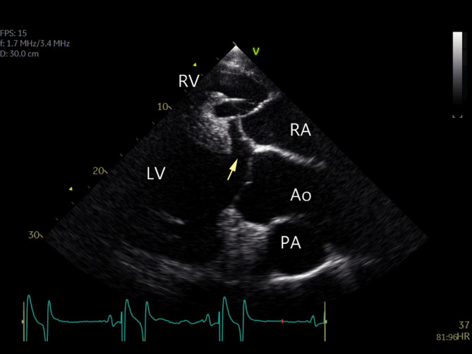
FIGURE 3.

An isolated perimembranous ventricular septal defect (arrow), viewed from a right parasternal left ventricular outflow image. There is no prolapse of the aortic valve. Ao, aorta; LV, left ventricle; PA, pulmonary artery; RA, right atrium; RV, right ventricle
