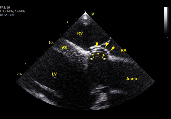
FIGURE 4.

A large isolated perimembranous ventricular septal defect (pmVSD), viewed from a right parasternal left ventricular outflow image in a 15‐year‐old warmblood horse. The marked bulging of the sinus of Valsalva (arrows) into the defect reduces the VSD shunt area but also causes aortic valve regurgitation. The double arrow shows the pmVSD longitudinal diameter. Arrowheads indicate the septal leaflet of the tricuspid valve. IVS, interventricular septum; LV, left ventricle; PA, pulmonary artery; RA, right atrium; RV, right ventricle
