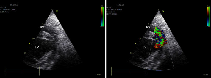
FIGURE 6.

A right parasternal short‐axis view near the ventricular apex (cranial to the right) without (left) and with (right) color flow Doppler. The small muscular ventricular septal defect is difficult to visualize on 2D because it is located ventrally and near the junction of septum and free wall. With color flow Doppler the systolic turbulent flow (green) into the right ventricle can be identified. LV, left ventricle; RV, right ventricle
