. 2020 Oct 21;11(48):12937–12954. doi: 10.1039/d0sc03245j

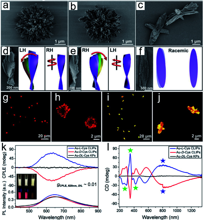
Fig. 3. Gold thiolate hierarchically organized particles (HOPs) with cysteine surface ligands.48 (a to c) SEM images of Au-l-Cys (a), Au-d-Cys (b) coccolith-like particles (CLIPs) and Au-dl-Cys (c) kayak particles with a low magnification. (a to c) Enlarged SEM images of Au-l-Cys (a), Au-d-Cys (b) CLIPs and Au-dl-Cys (c) kayak particles. (d to f) SEM images and corresponding schematic illustrations of segments of Au-l-Cys (d), Au-d-Cys (e) and Au-dl-Cys (f). Statistical analysis of SEM images for 100 assembly segments indicates that the pitch of individual nanoribbons in the stacks is 1300 ± 123 nm and their average width is 16 ± 1.8 nm, while the average angle between two neighboring nanoribbons in the stack is 7 ± 0.7° with the pitch of nanoribbon stacks being 820 ± 10 nm. (g to j) Confocal microscopy images of Au-l-Cys (g and h) CLIPs, and Au-dl-Cys (i and j) kayak particles. Chemical properties of Au-Cys dispersions in dimethylformamide and optical CLIPs. (k and l) CPLE (k) and CD (l) spectra of Au-l-Cys (blue), Au-d-Cys (red) CLIPs and Au-dl-Cys (black) kayak particles. Inset in (k): photos of Au-l-Cys, Au-d-Cys and Au-dl-Cys dispersions under daylight and UV light illumination. Adapted from ref. 48. Copyright© Copyright 2020 The American Association for the Advancement of Science.

Fig. 3