. 2020 Oct 22;11(48):13044–13051. doi: 10.1039/d0sc03166f

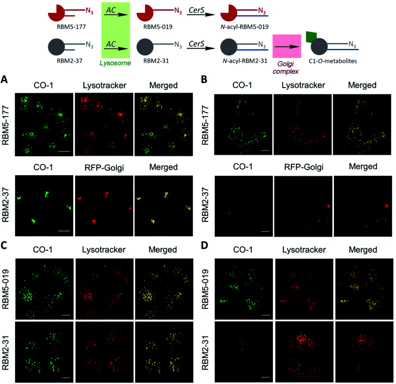
Fig. 3. Co-localization studies of the indicated probes in A549 cells. (A and C) A549 cells incubated or not with RFP-Golgi were treated with the indicated probe (5 μM, 20 min), washed and incubated with CO-1 (1 μM, 10 min). (B and D) After the probe treatment, cells were washed and incubated with medium for 5 h, washed and incubated with CO-1 (1 μM, 10 min). Lysotracker (75 nM) was added 30 minutes before fluorescence confocal microscopy (scale bar: 10 μm). Co-localization coefficients were measured on the different stacks of images (at least n = 4) with each stack containing 3–10 cells. Pearson's (P) and Manders' (M1 and M2) coefficients: (A) RBM5-177-Lysotracker (P = 0.802; M1 = 0.866; M2 = 0.744) and RBM2-37-RFP-Golgi (P = 0.615; M1 = 0.588; M2 = 0.804). (B) RBM5-177-Lysotracker (P = 0.746; M1 = 0.822; M2 = 0.711) and RBM2-37-RFP-Golgi (P = 0.346; M1 = 0.210; M2 = 0.206). (C) RBM5-019-Lysotracker (P = 0.713; M1 = 0.670; M2 = 0.774) and RBM2-31-Lysotracker (P = 0.731; M1 = 0.768; M2 = 0.570). (D) RBM5-019-Lysotracker (P = 0.762; M1 = 0.735; M2 = 0.688).

Fig. 3