. 2020 Jul 6;11(30):7910–7920. doi: 10.1039/d0sc01658f

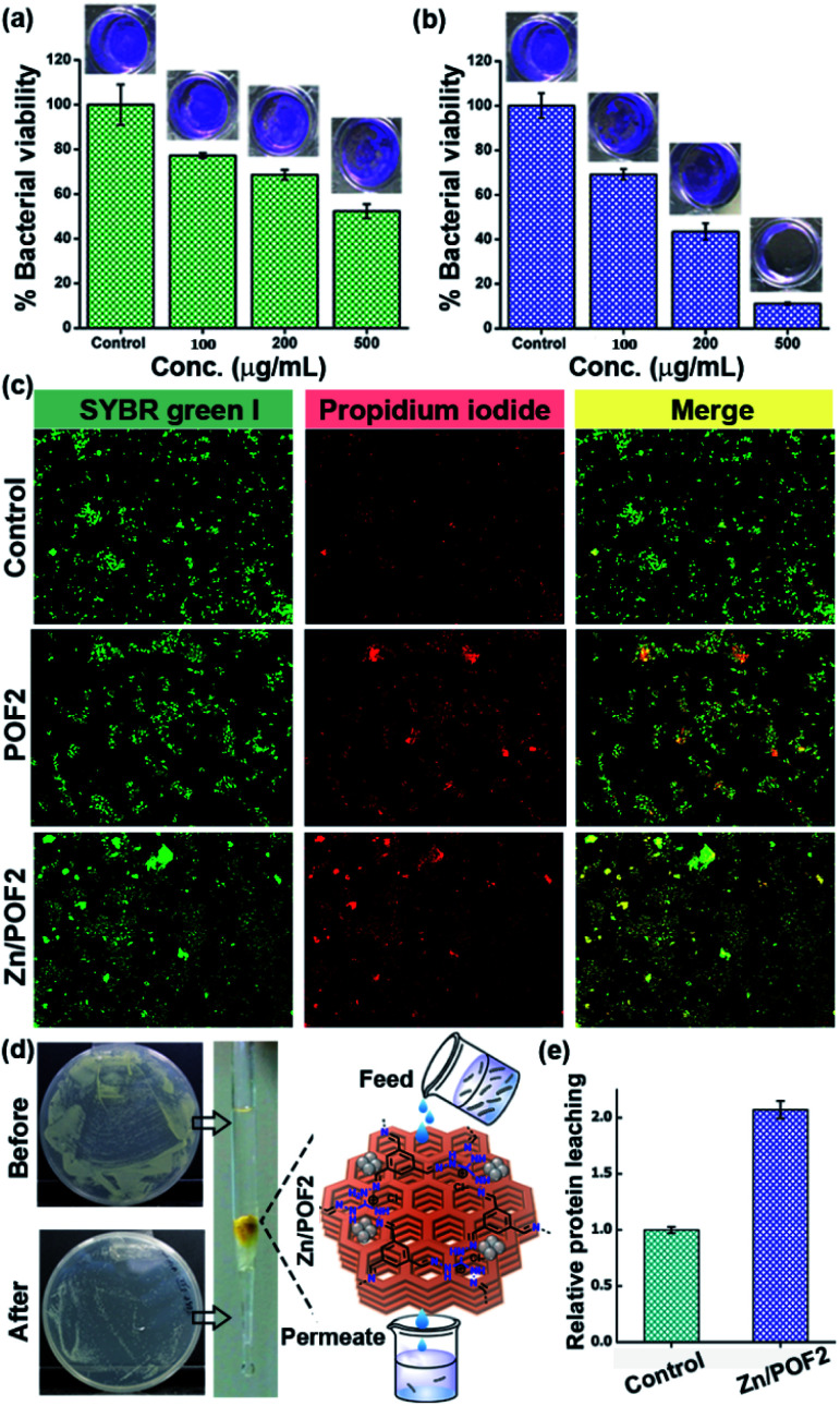
Fig. 5. The E. coli biofilm disruption due to various concentrations (μg mL−1) of (a) POF2 and (b) Zn/POF2. (c) Live/dead bacterial cell imaging using a dye mixture of SYBR Green I and propidium iodide staining of untreated (control), POF2, and Zn/POF2-treated E. coli cells. (d) The clearance of bacteria-contaminated water (E. coli ∼108 CFU mL−1) by passing through a column packed with the porous framework, Zn/POF2, evidenced by the CFU plating studies. (e) The relative protein leaching in the column flow-through using the Bradford assay, suggesting bacterial lysis after passing through the column of Zn/POF2. The Y-axis values were normalized to the eluted fraction obtained from the agarose-packed control column.

Fig. 5