. 2020 Jul 6;11(30):7910–7920. doi: 10.1039/d0sc01658f

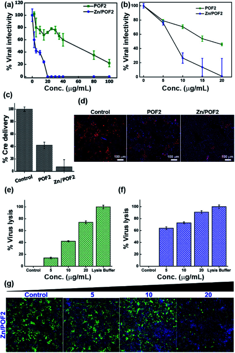
Fig. 6. The single cycle (a) human immunodeficiency virus-1 (HIV-1) and (b) vesicular stomatitis virus glycoprotein (VSV-G)-pseudotyped lentiviral vector infectivity in TZM-GFP target cells scored using a high-content screening platform and expressed in percentage with respect to the control; POF2 (green) and Zn/POF2 (blue). (c) The fusion assay of VSV-G lentiviral pseudoparticles against the porous frameworks POF2 and Zn/POF2 (20 μg mL−1). The error bars represent the standard deviation from the mean (n = 4, mean ± sd). (d) The representative images of Cre delivery as a function of fusion with the porous frameworks, POF2 and Zn/POF2 (20 μg mL−1), demonstrating the lower number of red fluorescent protein (RFP) positive cells in the case of Zn/POF2 than POF2 than control. The virus (VSV-G) lysis assay experiment showing the percentage of virus lysis in comparison with the lysis buffer against (e) POF2 and (f) Zn/POF2 with varying concentrations (n = 4, mean ± sd). (g) The assay demonstrating the loss of infectivity concomitant with virus lysis with the increasing dose (μg mL−1) of Zn/POF2; green cells represent the viral infected cell population, and Hoechst stained blue cells represent the total cell population.

Fig. 6