. 2020 Aug 12;11(33):8973–8980. doi: 10.1039/d0sc00929f

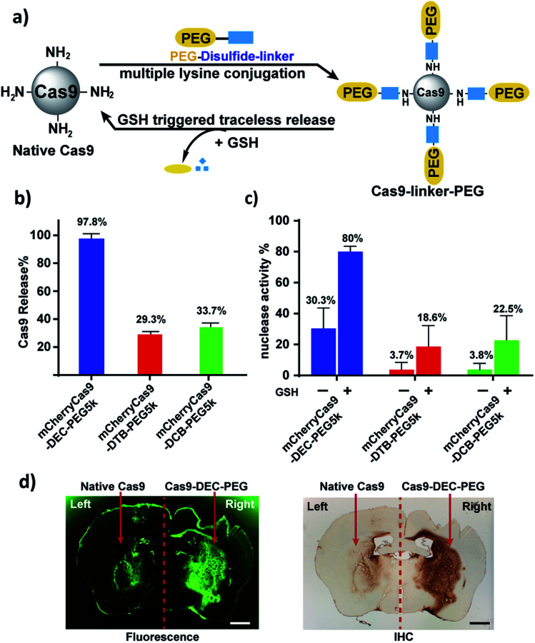
Fig. 3. mCherryCas9 can be conjugated to PEG via DEC and regains activity after reduction by GSH. (a) PEGylation of mCherryCas9 via three different disulfide linkers (DEC, DTB and DCB). (b) Release of PEG from mCherryCas9–disulfide–PEG after GSH (10 mm) incubation for 1 hour. mCherryCas9 release was analyzed by SDS-PAGE gel. (c) In vitro nuclease activity of the three mCherryCas9–disulfide–PEG conjugates was evaluated before and after reduction by GSH (10 mm). (d) Striatal distribution in the mouse brain after injection with Alexa488Cas9 (left striatum) or Alexa488Cas9–DEC–PEG5k (right striatum). Immunohistological staining (IHC) against Cas9 protein was visualized with DAB peroxidase substrate kit. Fluorescence microscope (green channel: 488 nm) scale bar: 1 mm.

Fig. 3