FIG. 3.
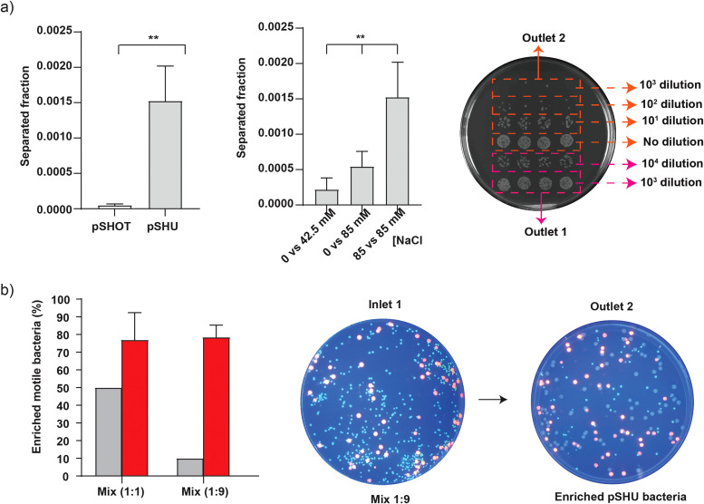
Separation and enrichment of sodium-motile bacteria. Inlet samples are configured as per Fig. 1. (a) Left—separation of motile pSHU bacteria and non-motile pSHOT bacteria, suspended in motility buffer containing 85 mM of NaCl and run against the solution of motility buffer containing 85 mM NaCl, Middle—selection of motile pSHU bacteria, suspended in 0 mM of NaCl vs varying concentration of sodium (42.5 and 85 mM of NaCl), and Right—droplet method to count bacterial colony collected from outlet 2 with no dilution, 10, and 102 dilutions whereas outlet 2 with 103 and 104 dilutions. (b) Left—% of enriched motile pSHU bacteria for two different mixtures of fluorescent cells in inlets (pSHU:pSHOT; 1:1 and 1:9), middle—spread plate of fluorescent cell mix ration of 1:9 (104 diluted) subjected into inlet 1 of microfluidic device, and right—spread plate of 100 μl collected cells from outlet 2 after separation (no dilution). Error bars represent standard deviation (SD) of triplicate data obtained from three independent experiments.

