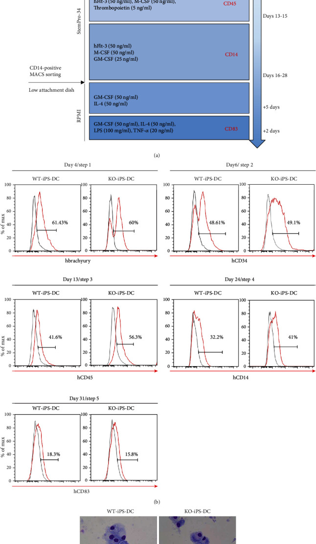
Figure 4.

Differentiation of dendritic cells derived from wild-type and HLA DR knockout iPSCs. (a) Schematic representation of differentiation protocol for iPSCs into dendritic cells. (b) iPS cell-derived dendritic cells on day 4 in the 1st step, day 6 in the 2nd step, day 13 in the 3rd step, day 24 in the 4th step, and day 31 in the 5th step were analyzed for the expression of brachyury, CD34, CD45, CD14, and CD83, respectively. (c) May-Grunwald-Giemsa staining of mature iPS induced DC on the glass slide is shown.
