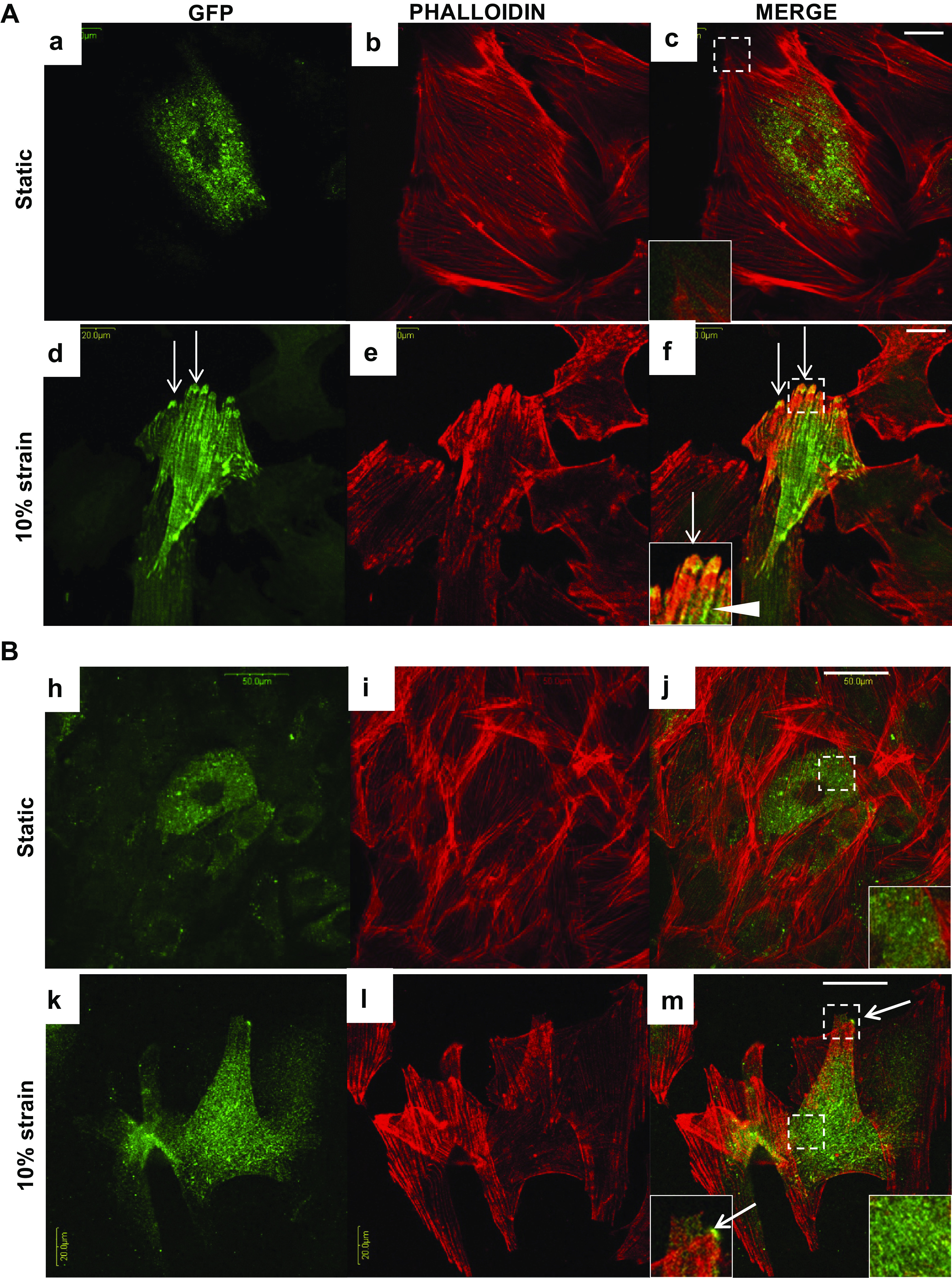
Figure 7.

Analysis of nebulette (NEBL)-green fluorescent protein (GFP) destribution in H9C2 cells at 24-h and 72-h under cyclic strain. Representative images of coimmunocytochemical analysis of GFP (green) and phalloidin (red) in H9C2 cells at 24-h (Aa–Ac and Bh–Bj, bar = 20 μm) and at 72-h (Ad–Af and Bk–Bm, bar = 50 μm) at the static or under strain conditions. Bottom left insets in the merged images represent focused detals of the squares outlining NEBL-GFP (arrows) at the focal adhesions, and bottom right insets represent NEBL-GFP expression in the cytosol.
