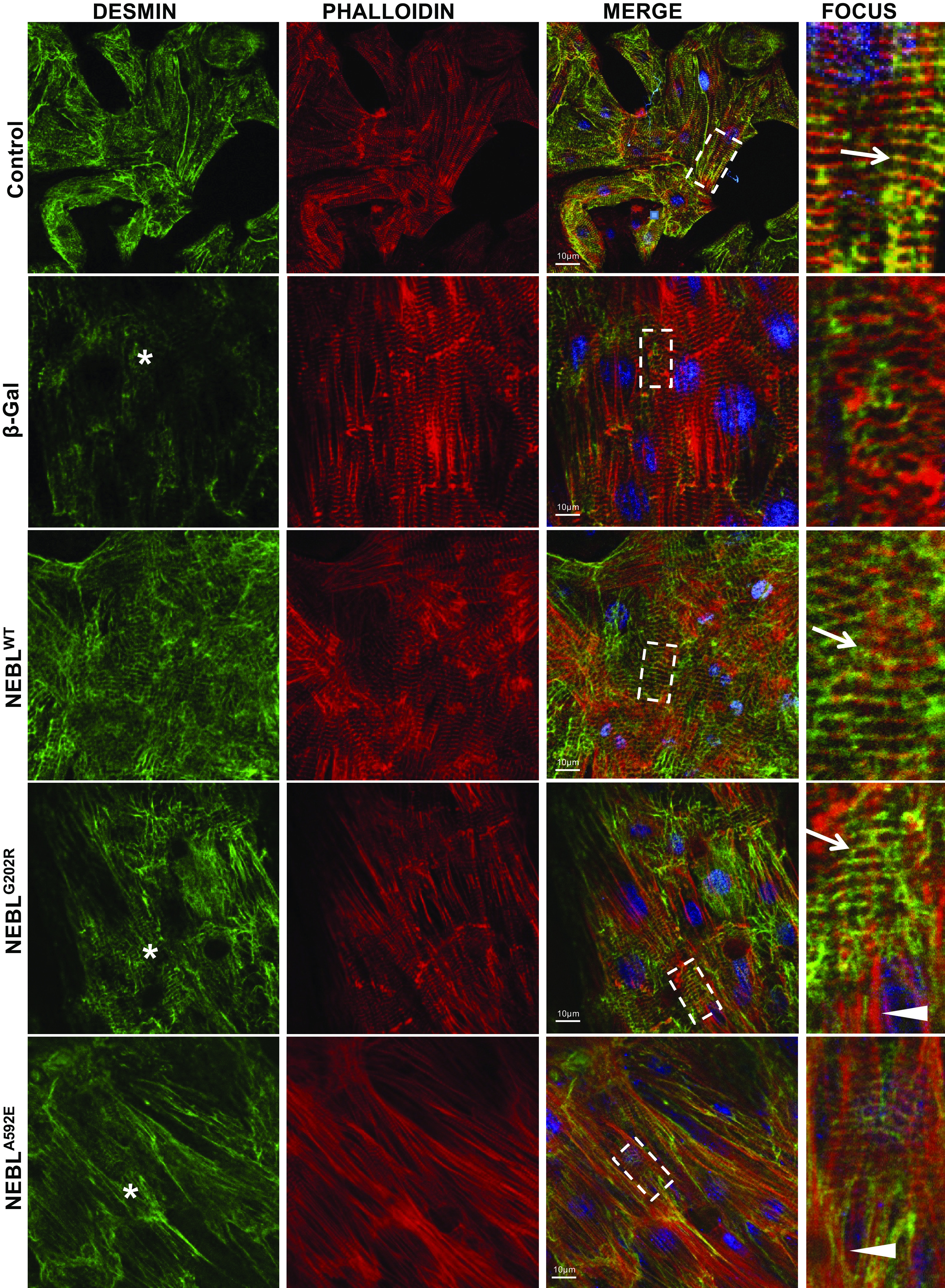
Figure 8.

Immunohistochemical analysis of desmin and actin structure in neonatal rat cardiomyocytes (NRCs) expressing wild-type (WT) or mutant nebulette (NEBL) under strain. Representative images of coimmunocytochemical analysis of desmin (green) and actin (phalloidin, red) in NRCs, uninfected (control), expressing β-gal, human NEBLWT, mutant NEBLG202R, or NEBLA592E delivered using adenoviral vectors under strain conditions. Scale bar = 10 μm. Fourth column: details of the square outlined in the merged images. Arrows indicate striation-like overlap of desmin and actin (yellow), and asterisks indicate disrupted desmin.
