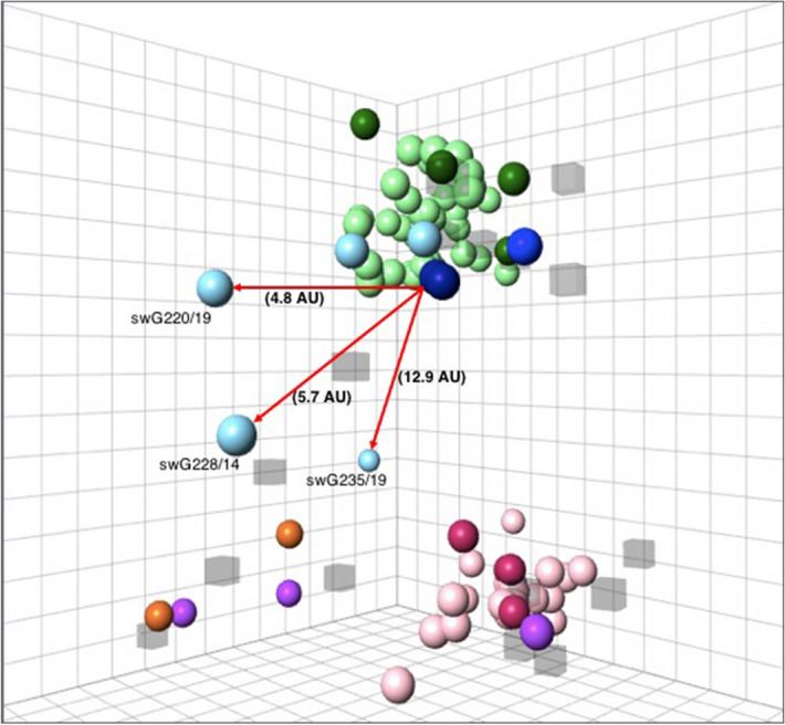
Figure 3.

Antigenic diversity of H1 swIAV isolated in Belgium and the Netherlands in 2014–2019. A 3D map was constructed based on the reactivity of the isolates in HI assay, with sera against the reference viruses, which included: dark green sphere EU H1av, maroon sphere EU H1hu, dark blue sphere H1pdm09, blue sphere US H1γ, orange sphere US H1δ1 and purple sphere Human H1. The H1 isolates are colored using lighter shades of the color of the reference viruses belonging to the same or related lineage: light green sphere EU H1av, pink sphere EU H1hu and light blue sphere H1pdm09. The virus strains are shown as spheres while the respective antisera are shown as semi-transparent cubes. Antigenic distances of the three H1pdm09 outliers from the H1pdm09 reference virus (CA09) are shown with red arrows. The squares in the grid represent antigenic distances. One square is equal to a two-fold change in HI titer, which is equal to 1 antigenic unit (AU). Analysis of the table vs map distance was conducted generating a moderately high r2 score value (r2 = 0.71).
