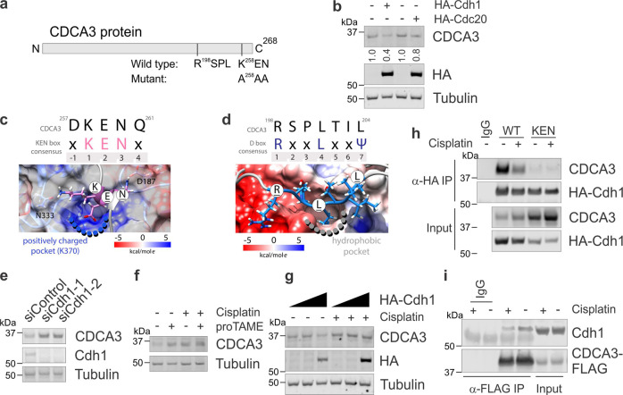
Fig. 3. CDCA3 avoids APC/CCdh1-mediated degradation following exposure to cisplatin.
a Schematic of primary CDCA3 protein sequence listing C-terminal D-box (RxxL) and KEN box motifs. KEN box mutated to A258AA also indicated. b Endogenous CDCA3 western blot analysis of H460 cells ectopically expressing increasing levels of HA-Cdh1 or HA-CDC20. Tubulin used as loading control and HA probe to determine ectopic Cdh1 or CDC20 expression. c, d In silico models of the CDCA3 KEN box (c) and D-box (d) bound to Cdh1. Snapshots were taken after 300 ns of molecular dynamics simulation. Upper, CDCA3 protein sequence compared with consensus motifs. X = any amino acid, Ψ = aliphatic amino acid. Numbers indicate amino acid position within motif. Lower, colour scales highlighting the Columbic electrostatic potential at the surface of Cdh1 in these functional regions. e Western blot analysis of endogenous CDCA3 in control or Cdh1 depleted H460 cells. f Endogenous CDCA3 western blot analysis of H460 cells treated with cisplatin, the APC/C inhibitor proTAME or combination of both. g Western blot analysis of H460 cells expressing increasing levels of ectopic HA-Cdh1, as per Fig. 3b, treated with or without cisplatin. h Immunoprecipitation analysis of ectopic HA-Cdh1 with wild-type or KEN mutant CDCA3-FLAG from H460 cells treated with or without cisplatin. i Reciprocal immunoprecipitation analysis of ectopic CDCA3-FLAG with endogenous Cdh1 in H460 cells treated with or without cisplatin. All in vitro experiments are representative of three independent repeats.

