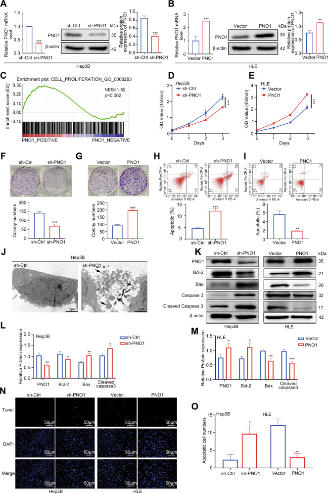
Fig. 2. PNO1 promotes HCC cell proliferation and inhibits cell apoptosis.
A, B The construction of Hep3B PNO1- downregulation (sh-PNO1) and HLE PNO1-upregulation (PNO1) cell. C GSEA validating the correlation between PNO1 expression and the cell proliferation genes. D, E CCK-8 assay analysis of the impact of PNO1 knockdown or overexpression on Hep3B and HLE cell growth. F, G Colony formation assay showing the effects of PNO1 knockdown (F) or overexpression (G) on Hep3B and HLE cell growth. H, I Apoptotic rate was measured using annexin V/PI double staining in PNO1 knockdown (H) or overexpression (I) cells. J TEM analysis of cell ultrastructural characteristics in Hep3B sh-PNO1 and sh-Ctrl cells. Chromatin condensation and nuclear fragmentation are indicated by arrows. Scale bar = 20 μm. K Western blotting analysis of apoptosis-related protein levels in cells as in (A) and (B). L, M Quantification of relative protein expression. N, O Apoptosis was also evaluated by TUNEL staining (N) and quantification of TUNEL positive cells (O). Scale bar: 50 μm. Data were presented as mean ± SEM. n = 3–4. *p < 0.05, **p < 0.01, ***p < 0.001.

