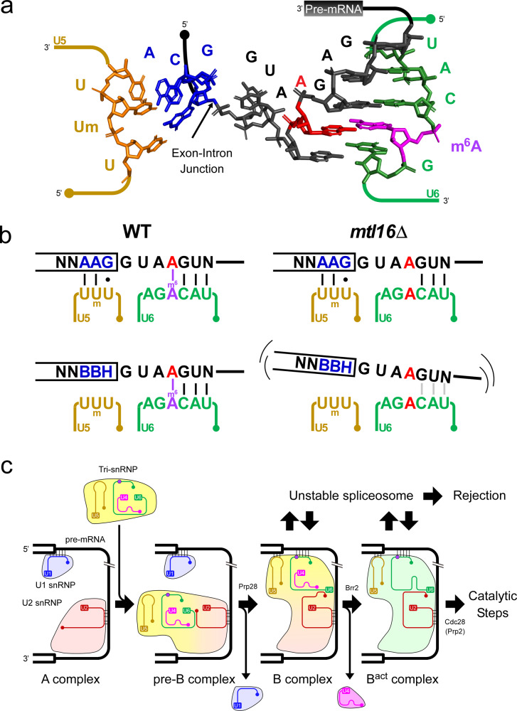
Fig. 5. Mechanistic insight into the functional role of U6 snRNA m6A modification on pre-mRNA splicing.
a Cryo-EM structure of 5′SS in the B complex of the human spliceosome (PDB ID:5O9Z)92, as depicted by PyMol 2.1.0 (http://www.pymol.org/2/support.html). The model consists of U5 snRNA (orange), U6 snRNA (green), 5′ exon (blue), intron (black), A4 (red), and m6A (magenta). The coordinate of the methyl group of m6A was set to its syn conformation. b Interaction between the 5′SS of A4 intron and the U5 and U6 snRNAs in WT and mtl16Δ strains. m6A–A pairing stabilizes the complex, especially when U5 snRNA recognizes the 5′ exon weakly. c Schematic depiction of spliceosomal assembly from A to Bact complex based on cryo-EM structures of S. cerevisiae and human spliceosomes83–86,91,92. The m6A base in U6 snRNA is depicted by a dark-purple circle. The blue, red, yellow, purple, and green figures represent U1 snRNP, U2 snRNP, U4/U6.U5 tri-snRNP, U4 snRNP, and Bact complex, respectively.

