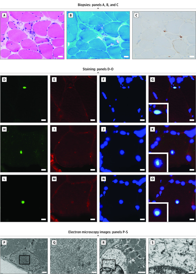
Figure 4. Histopathologic and Electron Microscopy Findings in Patients With OPDM_LRP12 Patients.
A, B, and C, Biopsies from the left biceps brachii of patient 21, who has had the disease for 8 years, are shown. Scale bar: 20 μm. A, Moderate to marked fiber size variation and fibers with internal nuclei are seen on hematoxylin-eosin stain. B, Fibers with rimmed vacuole and moderate fibrous tissue infiltration are seen on modified Gomori trichrome stain. C, The dotlike deposition of p62 can be observed in muscle fibers. Staining with anti–SUMO-1 antibody (D), anti–caveolin-3 antibody (E, I, M), 4′,6-diamidino-2-phenylindole (F, J, N), anti-p62 (H), and anti–poly-ubiquitinated protein antibody (L). G, K, and O show merged immunohistochemistry. D to O, Scale bar: 10 μm. P and Q, Electron microscopy images of the biopsied left biceps brachii muscle from patient 10, in their late 60s. Scale bars are 1 μm for P and 200 nm for Q: Intranuclear tubulofilamentous inclusions (mean [SD] diameter, 17.3 [1.4] nm) are shown. R and S, Electron microscopy images of the biopsied left deltoid from patient 14, in their late 30s. Scale bars are 1 μm for R and 200 nm for S: Cytoplasmic tubulofilamentous inclusions (diameter, 10 nm) are shown.

