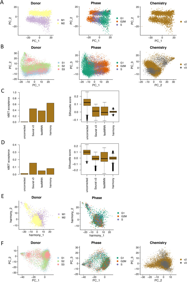
Fig. 2.
Batch effects and batch-effect correction. a Dimensional reduction plot on the first two principal components showing the distribution of menstrual ePCs color coded by donor, cell cycle phase, and chemistry reagent version. Clear donor effect and cell cycle effect were observed. b Dimensional reduction plot on the first three principal components showing the distribution of secretory ePCs color coded by donor, cell cycle phase, and chemistry reagent version. Clear donor effect, cell cycle effect, and chemistry reagent version effect were observed. c Evaluation on batch-effect correction methods using kBET acceptance score (left panel) and Silhouette score (right panel) on menstrual ePCs. All three methods Seurat v3, fastMNN, and harmony removed the batch effects to different extents when compared to the uncorrected data. Harmony ranked the best by achieving the highest kBET acceptance score and narrowest Silhouette score. d Evaluation on batch-effect correction methods using kBET acceptance score (left panel) and Silhouette score (right panel) on secretory ePCs. All three methods Seurat v3, fastMNN and harmony removed the batch effects to different extents when compared to the uncorrected data. Seurat v3 ranked the best by achieving the highest kBET acceptance score and lowest Silhouette score. e Dimensional reduction plot on the first two harmony dimensions showing the distribution of cells color coded by donor after batch effect correction and cell cycle phase after cell cycle regression. f Dimensional reduction plot on the first two principal components showing the distribution of cells labeled by donor after batch effect correction and cell cycle phase after cell cycle regression

