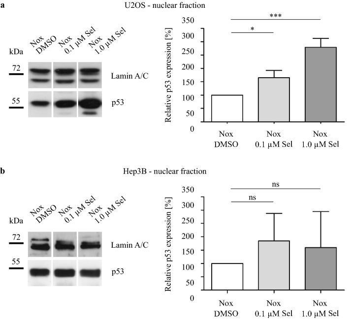
Fig. 2.
Immunoblot analysis of intranuclear p53 protein level. a) U2OS and b) Hep3B cells were treated with DMSO, 0.1 µM Selinexor or 1.0 µM Selinexor under normoxia for 24 h and fractionated. Hep3B cells are deficient for the tumor suppressor protein p53 and were transiently transfected with a p53-pcDNA-plasmid. Relative protein levels of intranuclear p53 protein in % ± SD compared to DMSO control are shown. One-way ANOVA with Tukey posttest with *p < 0.05 and ***p < 0.001 (n = 3)

