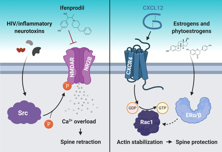
Fig. 1.
Dendritic spine loss in HAND models can be reversed by different approaches. Recent studies have reported possible approaches to reverse dendritic spine deficits in animal models of HAND, which also improved their cognitive function. These approaches involved inhibiting excess Ca2+ influx into neurons (left panel) and promoting neuronal actin stabilization (right panel). In the left panel, HIV neurotoxin activation of Src causes excessive Ca2+ influx through NR2B-containing NMDA receptors (NMDAR) in the retrosplenial cortex, but inhibiting these receptors prevents excessive Ca2+ influx and spine retraction in the same region. In the right panel, CXCL12 binding to CXCR4 on cortical neurons activates a Rac1-mediated pathway that stabilizes actin, a critical structural component of dendritic spines, which protects existing spines. Interestingly, CXCL12 is also able to downregulate NR2B-containing NMDARs. Moreover, various estrogen and phytoestrogen molecules can increase dendritic spine density via estrogen receptor (ERα/β) signaling, which may also involve actin stabilization via Rac1 signaling. The mechanisms presented in this figure are explained in detail in the next section

