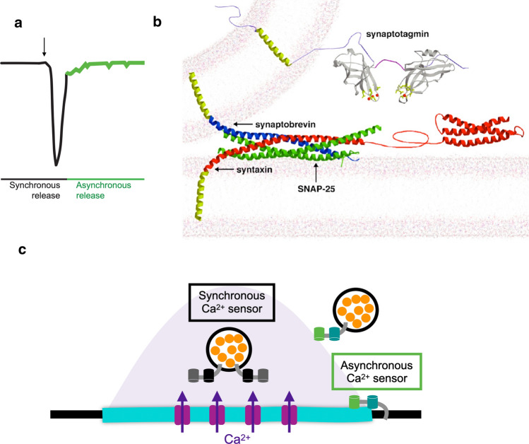
Fig. 1.
Regulation of synchronous and asynchronous SV fusion. a Model depicting phases of synchronous and asynchronous release after nerve stimulation (arrow). b Structure of the fusion machinery that bridges the SV and plasma membrane, with SYT1 (grey) and the SNARE complex (Syntaxin—red; SNAP25—green; Synaptobrevin—blue). c Model of synchronous and asynchronous release in relation to presynaptic Ca2+ entry (shaded). Following Ca2+ influx, SVs fuse during a synchronous phase at AZs that occurs within milliseconds. SYT1 acts as the Ca2+ sensor for synchronous release and resides on SVs. A slower asynchronous component can last for hundreds of milliseconds and include fusion of SVs farther from release sites. SYT7 has emerged as a candidate for the asynchronous Ca2+ sensor with higher affinity than SYT1 for Ca2+ binding. The role of SYT7 is still controversial, with studies in Drosophila suggesting it controls SV availability and fusogenicity

