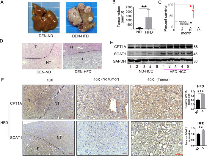
Fig. 1. HFD improves the expression of SOAT1 and CPT1A in DEN-induced HCC.
A–B A high-fat diet exacerbated HCC progression in DEN-injected mice. Representative images of the livers (A) and tumor volumes (B), ND: the normal diet group (n = 8) and HFD: the high-fat diet group (n = 5). (mean ± SEM) **p < 0.01. C Kaplan–Meier survival plot of DEN-injected mice on a high-fat diet (HFD) or normal diet (ND); *p < 0.05. D H&E-stained images of livers using HCC and adjacent non-tumor liver tissue in ND-fed or HFD-fed mice (scale bar, 50 μm). NT and T indicated non-tumor and tumor areas, respectively. E Western blot analysis of SOAT1 and CPT1A in liver cancer tissues from ND-fed or HFD-fed mice. F Representative IHC staining (n = 5 images in total) of SOAT1 or CPT1A in the tissues obtained from HFD-fed mice. NT and T indicate non-tumor and tumor areas, respectively (scale bar, 50 μm). Five separate areas from each tissue were quantified (mean ± SEM); *p < 0.05; **p < 0.01, ***p < 0.001.

