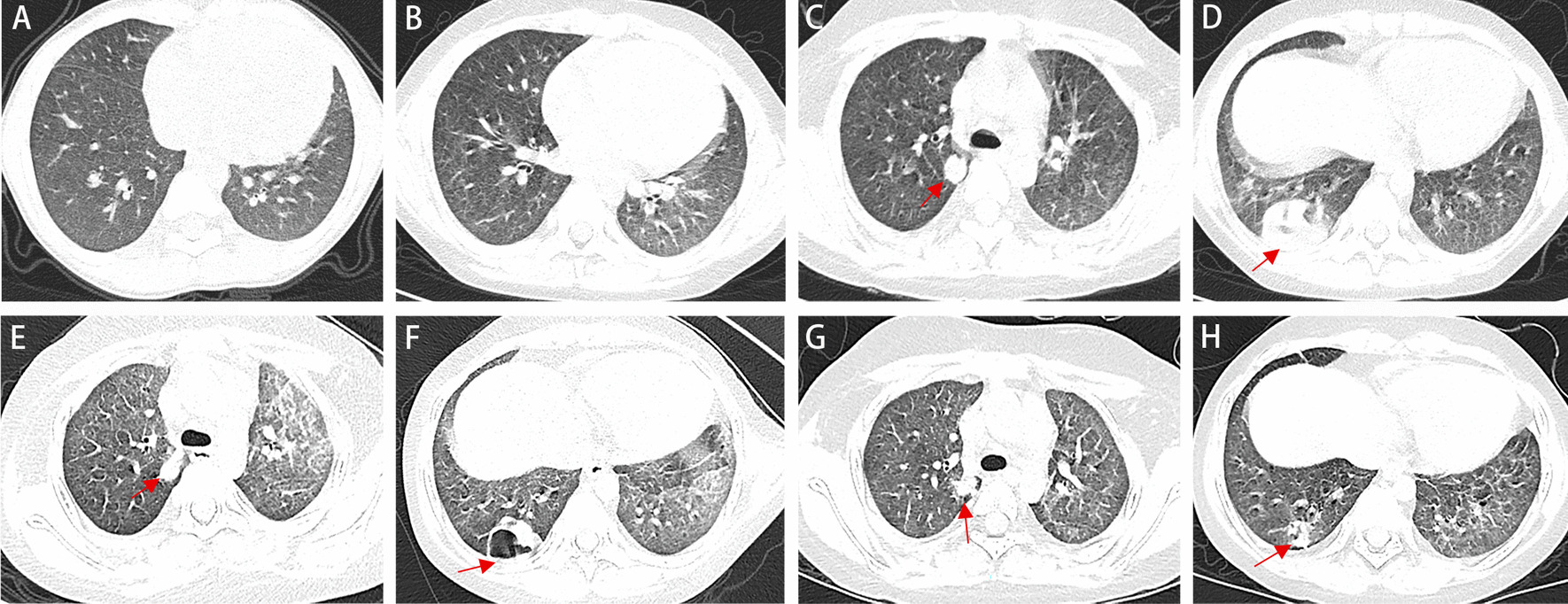
Fig. 1.

Changes in chest imaging of the patient. a Diffuse fine granular shadows were observed in both lungs (2 months before admission). b Diffuse granular shadows in both lungs and round nodular high-density shadows in the right lung (14 days before admission). c–d The transmittance of the disease into the bilateral lung decreased unevenly, and there were still fine particles in the lungs. The right lung contained scattered, circular lesions, and the boundaries were clear, the arrors showed two large lesions, the larger is about 30 × 42 × 27 mm (on the day of admission). e, f The lesions in the lower lobe of the right lung were improved, but cavity formation, new lesions in the left lung, and bilateral pleural thickening were observed (on the 9th day of admission). g, h The lesions in the inferior lobe of right lung with cavity were significantly reduced, and the changes of both lungs were improved (on the 16th day of discharge)
