Figure 1.

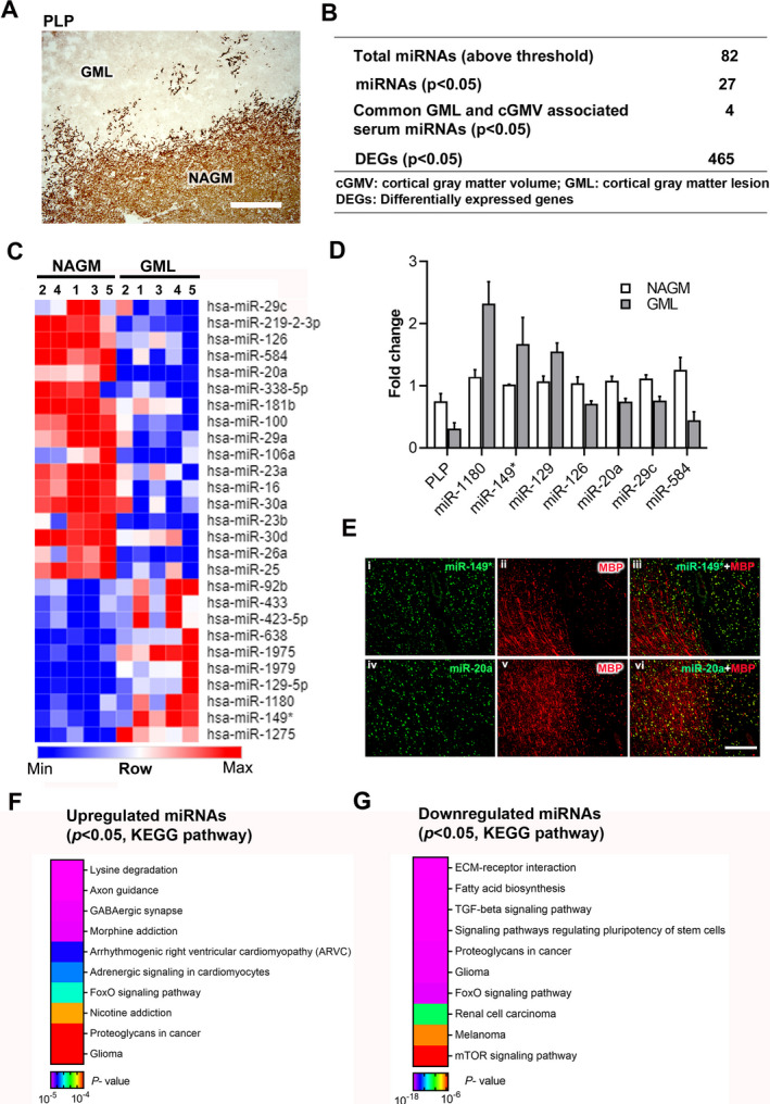
miRNAs are dysregulated in cortical demyelinated lesions. (A) Representative immunohistochemical staining of proteolipid protein (PLP) showing a demyelinated GML and surrounding NAGM from a progressive MS brain. Scale bar—200 µm. (B) Summary table of microarray miRNAs and gene expression analysis. (C) Heatmap image shows significantly upregulated and downregulated miRNAs identified in GMLs. (D) qPCR validation analysis of PLP and selected miRNA expression in NAGM and GMLs from progressive MS brains (n = 3–4). (E) Immuno‐in situ hybridization (immuno‐in situ) of miRNAs (green) and myelin protein (MBP, red) showing miR‐149* (i–iii) and miR‐20a (iv–vi) in myelinated and demyelinated lesioned areas of progressive MS brain. miR‐149* showed increased expression in lesioned areas, whereas miR‐20a was decreased in GMLs. GML, gray matter lesion; NAGM, normal‐appearing gray matter. (F, G) Representative heatmap image showing KEGG pathways of genes targeted by (F) upregulated and (G) downregulated miRNAs in GMLs.
