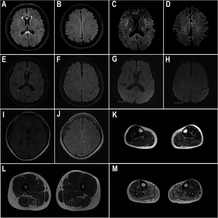
Figure 2.

Brain and muscle MRI findings. (A‐J) Brain MRIs of F1‐III‐5 (A‐D), S1 (E‐H), and F2‐IV‐5 (I, J). (A, B, E, F) showed mildly high signal on the splenium of corpus callosum on T2‐FLAIR. (C, D) showed no obvious abnormalities on DWI. (G‐J) showed no obvious abnormalities on DWI (G, H) or T2‐FLAIR (I, J). (K‐M) Muscle MRIs on T2‐FLAIR were evaluated at 8 and 4 years after the onset of symptoms in patients F2‐IV‐5 (K) and F1‐III‐5 (L, M), respectively. Remarkable fatty degeneration and mild amyotrophy were observed. The distal muscles (M, calf level) were more severely affected than the proximal muscles (L, thigh level).
