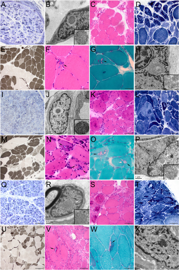
Figure 3.

Pathological changes of nerve and muscle biopsies from patients F1‐III‐5 (A‐H), F2‐IV‐5 (I‐P), and S1 (Q‐X). (A, I, Q) Toluidine blue staining of nerve sections showed a reduced density of myelinated nerve fibers. A few thin‐myelinated nerve fibers were present. Ultrastructural examination of peripheral nerve tissues (B, J, R) demonstrated that the intranuclear inclusions containing filamentous aggregates (B, fibroblast; J, capillary pericyte; R, Schwann cell). (C‐G, K‐O, S‐W) Muscle biopsies showed both neurogenic changes (groups of small angular atrophic muscle fibers, type grouping, and target or targetoid fibers) and myopathic changes (rimmed vacuoles fibers, swirling fibers, endomysial fibrosis). (H, P, X) Ultrastructural examination of muscle tissues demonstrated that the intranuclear inclusions containing filamentous aggregates. (C, F, K, N, S, V) H&E staining; (D, L, T) NADH staining; (E, M, U) ATPase staining (pH 10.7, pH 4.4, pH 10.5, respectively); (G, O, W) mGT staining. Scale bars = 50 μm in A, D, F, G, I, N, O, Q, T, W; 100 μm in C, E, K, L, M, S, V; 200 μm in U; 1 μm in H, J, P; 0.5 μm in B, R, X.
