
Figure 5.
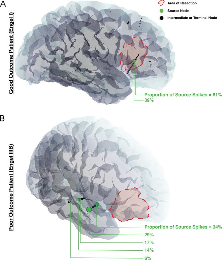
Patient‐specific imaging with an overlay of network nodes. Only the SEEG electrode contacts involved in the patient's spike propagation network are shown; spiking regions that failed to demonstrate statistically significant propagation patterns are not displayed. (A) Seizure‐free patient (Engel I) with two source nodes, both included in the resection, for a total source spike concordance value of 100%. (B) Seizure‐persistent patient (Engel IIIB) with five source nodes, none being included in the resection, for a total source spike concordance value of 0%. Interictal spikes were present in the frontal lobe, but they did not demonstrate statistically significant propagation patterns