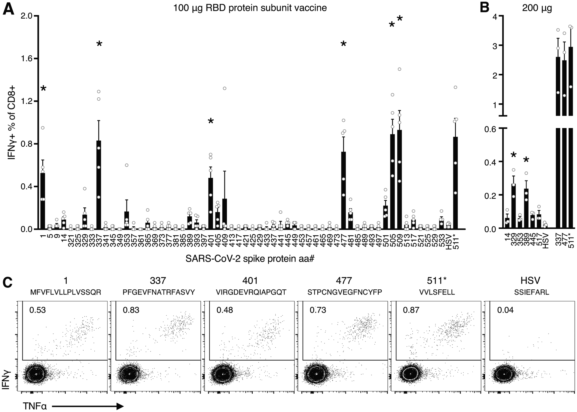
Figure 1. Epitope mapping of CD8 T cell responses to SARS-CoV-2 RBD protein in C57BL/6 mice.

Five mice were immunized with RBD protein plus adjuvant and their spleens harvested one week later. A) The percentage of CD8 T cells staining for IFNγ after a 5 h incubation with individual 15-mer peptides spanning SARS-CoV-2 RBD. Responses that were significantly greater than those induced by an irrelevant peptide (HSVgB498–504), as determined by Dunnett’s multiple comparisons test (where p<0.01), were indicated by an asterisk. B) The percentage of CD8 T cells staining for IFNγ for the six potential minor epitopes and three of the major epitopes identified in A) in mice immunized 200 μg of RBD plus adjuvant. C) Representative intracellular IFNγ and TNFα staining. Cells were pre-gated on lymphocytes, singlets, live cells, and CD8+CD4−B220−.
