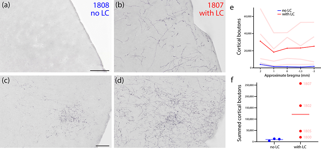
Figure 13.
Cortical labeling in cases with or without LC contamination. (a) The somatosensory cortex in case 1808 (no transduction of LC neurons) contained no Syp-mCherry labeling, while (b) the same region in case 1807 (many Syp-mCherry-expressing neurons in LC) had a uniform meshwork of Syp-mCherry labeling, similar to the rest of the cerebral cortex. The insular cortex in case 1808 (c) contained a moderate terminal field of grainy boutons, while case 1807 (d) had a similar terminal field, plus superimposed, uniform labeling similar to the rest of the cerebral cortex. (e) Quantification of all Syp-mCherry-labeled boutons across cases at 5 rostro-caudal levels of the cerebral cortex (approximate bregma levels shown along the x-axis). Pale red (LC-transduced) and blue (no LC) lines represent individual cases. Bold red and blue lines represent the average of cases from each group. (f) Summed cortical boutons (across all five counted brain sections) in each case.

