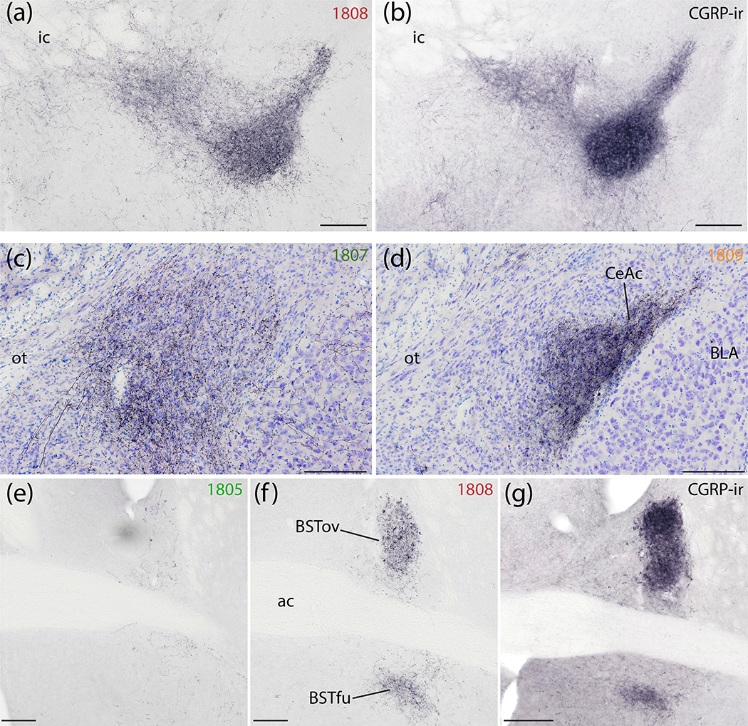
Figure 14.
Syp-mCherry labeling in the central nucleus of the amygdala (CeA) and bed nucleus of the stria terminalis (BST). (a) NiDAB immunolabeling for Syp-mCherry in the SI and rostral capsular CeA (case 1808) is strikingly similar to (b) CGRP immunolabeling in the same region of an uninjected C57B6/J mouse brain. (c) Syp-mCherry labeling was more prominent in the medial CeA subdivision after a more medial injection site in the PB region (case 1807) and (d) even more dense in the capsular and lateral subdivisions (CeAl and CeAc) following a more lateral injection, in PBeL (case 1809). (e–g) Labeling in the BST followed a topographical organization similar to the CeA. (e) Minimal labeling in the BST after a medial injection site (case 1805). (f) Denser Syp-mCherry labeling in the oval and fusiform subnuclei (BSTov and BSTfu) after a more lateral injection involving PBeL is strikingly similar to (g) CGRP immunolabeling in the same region of a an uninjected C57B6/J mouse brain. Scale bars are 200 μm (a–d) or 500 μm (e–g).

