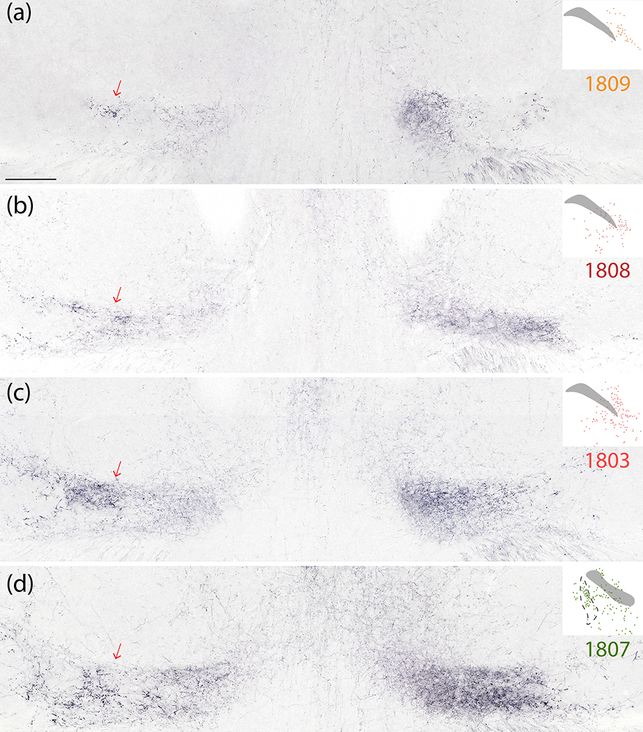
Figure 15.
Syp-mCherry labeling in the thalamic ventral posterior parvicellular nucleus (VPpc) was lighter after injections into the PBeL (case 1809, a) and progressively denser after injections involving more of the medial PB (cases 1808–1803-1807; b–d). Note that the lateral aspect of the VPpc contained large, bulky boutons which were more prominent on the contralateral side (red arrows at left). The medial aspect of the VPpc contained smaller, grainy boutons which were denser on the ipsilateral (right) side in every case. Inset at upper-right of each panel shows the distribution of Syp-mCherry-transduced neurons at the center of the injection site for each case. Scale bar is 500 μm (a) and applies to (b–d).

