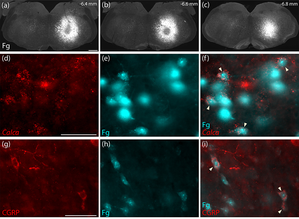
Figure 16.
Retrograde confirmation that PB CGRP/Calca neurons project axons to the hindbrain reticular formation (a–c) Rostrocaudal extent of Fluorogold (Fg) injection into the medullary reticular formation (case 3621). This injection site was centered above to the facial motor nucleus at rostrocaudal levels between the caudal cochlear nucleus and rostral inferior olivary nucleus. (d–f) Neurons in the rostral, ventral PB contain both Calca mRNA (red in d) and Fg (ice-blue in e–f). (g–i) In the same region, CGRP immunofluorescence labeling (red, in g) co-localized with Fg (ice-blue in h-i). Scale bars are 500 μm (a) and 50 μm in (d,g) and apply to the remaining panels in each row.

