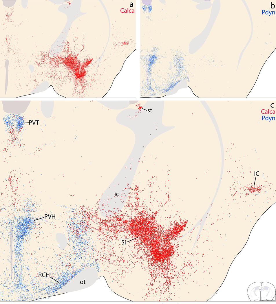
Figure 21.
Complementary Calca (red) and Pdyn (blue) patterns in the basal forebrain, hypothalamus, rostral thalamus, and insular cortex. (a) Calca projections densely innervate the substantia innominata (SI), with lighter labeling extending dorsally into the ventral fringe of the internal capsule (ic), globus pallidus, and between the striatum and pallidum. Lateral to the SI, the insular cortex (IC) contains light labeling. Medial to the SI, the hypothalamus has very little labeling. (b) Pdyn projections densely target the paraventricular nucleus of the hypothalamus (PVH) and the retrochiasmatic area (RCA) above the optic tract. In the thalamus, Pdyn projections concentrate beneath the third ventricle, in the dorsal aspect of the paraventricular nucleus of the thalamus (PVT). (c) Combined labeling.

