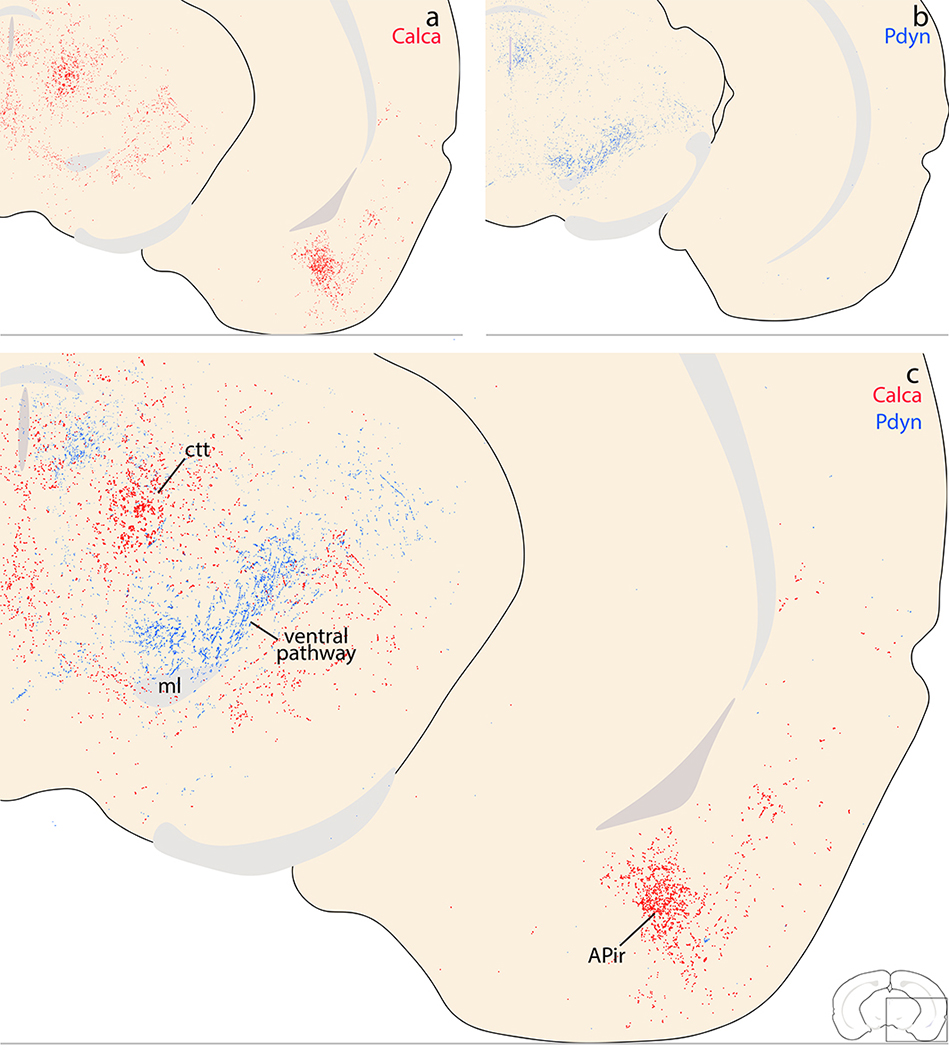
Figure 24.
Complementary Calca (red) and Pdyn (blue) patterns in the rostral midbrain. (a) The majority of Calca axons project through the central tegmental tract (ctt), ventrolateral to the periaqueductal gray matter (PAG). Calca projections to the PAG and other midbrain regions are light and scattered, but in the ventral cerebral cortex, a moderately dense terminal field appears in the amygdalopiriform transition area (APir). (b) Pdyn axons project through and form a light terminal field along the dorsolateral fringe of the medial lemniscus (ventral pathway). (c) Combined labeling shows that Calca axons projecting through the ctt are separate from Pdyn axons projecting through the ventral pathway.

