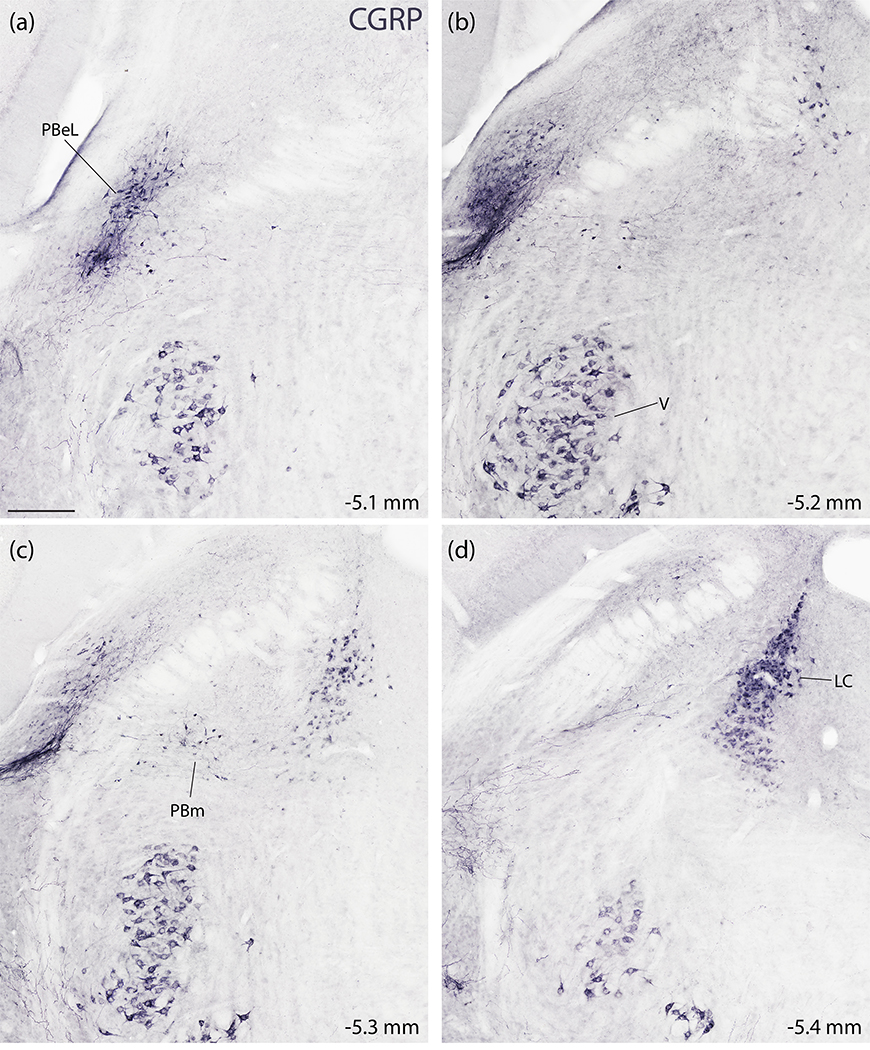
Figure 7.
Immunohistochemical labeling for CGRP in the PB region. (a–d) NiDAB immunohistochemical staining across four rostral (a) to caudal (d) levels of the PB region. CGRP immunolabeling is strikingly similar to the distribution of Calca mRNA-expressing neurons shown in figure 5. CGRP-immunoreactive neurons concentrate in the LC and the PBeL, with fewer in PBm and more in V (also VCo, not shown). Approximate distance caudal to bregma is shown at the bottom-right of each panel. Scale bar in (a) is 200 μm and applies to all panels.

